
SUPPORT AND MOVEMENTS
FILL IN THE BLANKS
Q.01: Fill in the blanks:
(i) Each muscle is enclosed by a membrane known as ______. (sarcolemma)
(ii) Osteoporosis in caused by the decrease in the level of ______. (estrogen)
(iii) The “molting” is controlled by a hormone ______. (ecdysone)
(iv) ______ is stored in the muscle cell as reserve food. (glycogen)
(v) Collenchymatous cells lack ______ in their primary wall. (secondary wall)
(vi) There are _____ vertebrae in the neck region of mammals. (seven)
(vii) The most abundant proteins in the muscle are ______. (actin & myosin)
(viii) ______ connect a muscle to a bone. (tendons)
(ix) Thick muscle filament is composed of ______. (myosin)
TRUE/FALSE
Q.02: Indicate True or False.
(i) The shoulder joint is a hinge joint. (FALSE)
CORRECT: The shoulder joint is a ball and socket joint.
(ii) Tendons connect bones together at joints. (FALSE)
CORRECT: Ligaments connect bones together at joints.
(iii) Arthritis often accompanies aging. (TRUE)
(iv) Calcium provides energy to the muscle contraction. (FALSE)
CORRECT: ATP provides energy to the muscle contraction.
(v) Most of the sclerenchymatous cells are non-living. (TRUE)
(vi) Visceral muscles are striated, involuntary and smooth. (FALSE)
CORRECT: Visceral muscles are non-striated, involuntary and smooth.
MULTIPLE CHOICE QUESTIONS
Q.03: Each question has four options. Encircle the correct option.
(i) Which of these is a direct source of energy for muscle contraction?
(a) ATP
(b) Creatine phosphate
(c) Lactic acid
(d) Both a and b
ANSWER: (a) ATP
EXPLANATION: ATP (adenosine triphosphate) serves as the immediate energy source for muscle contraction. When muscles contract, ATP binds to myosin, allowing it to detach from actin. The hydrolysis of ATP into ADP & inorganic phosphate provides the energy needed for myosin to reattach to actin, promoting the sliding of filaments and muscle contraction.
(ii) When muscle contracts:
(a) Sarcomere increases in size
(b) Myosin slides past actin
(c) Lactic acid is produced
(d) Both a and b
ANSWER: (b) Myosin slides past actin
EXPLANATION: During muscle contraction, myosin filaments generate force and slide past actin filaments. This sliding action is facilitated by the formation and breaking of cross-bridges between myosin and actin, powered by the energy released from ATP hydrolysis. As these filaments slide, the muscle shortens, leading to contraction.
(iii) Which of the following changes occurs when skeletal muscle contracts.
(a) The A band shortens
(b) The I-band shortens
(c) The Z-line slides farther apart
(d) The actin filament contracts
ANSWER: (b) The I-band shortens
EXPLANATION: During skeletal muscle contraction, the I-band shortens as actin filaments slide over myosin filaments, bringing the Z-lines closer together.
(iv) Thin filament in myofibrils consist of:
(a) Actin, tropomyosin, troponin
(b) Z-line
(c) Myosin
(d) Sarcomere
ANSWER: (a) Actin, tropomyosin, troponin
EXPLANATION: The thin filament in myofibrils is composed of actin, which forms the primary structure, while tropomyosin lies along the groove of the actin helix. Troponin is bound to tropomyosin and regulates muscle contraction by controlling the interaction between actin and myosin in response to calcium ions.
(v) The contraction of striated muscle is initiated by release of energy in the presence of:
(a) Acetyl choline
(b) Calcium ion
(c) Chloride ion
(d) Iron
ANSWER: (b) Calcium ion
EXPLANATION: The contraction of striated muscle is initiated by the release of energy when calcium ions bind to troponin, causing a conformational change that allows the myosin heads to bind to actin filaments and initiate the sliding filament mechanism, leading to muscle contraction.
(vi) In the mammalian skeleton, there is a distinct synovial joint between the:
(a) Bones of the cranium
(b) Humerus of ulna
(c) Sacrum of ilium
(d) Sternum and floating ribs
ANSWER: (b) Humerus of ulna
EXPLANATION: The synovial joint between the humerus and ulna is a typical example of a hinge joint found in the mammalian skeleton. This joint allows flexion and extension movements, enabling the bending and straightening of the arm.
(vii) Which of the following is a bone of axial skeleton?
(a) Rib
(b) Shoulder girdle
(c) Pelvis
(d) femur
(e) All of the above
ANSWER: (a) Rib
EXPLANATION: The axial skeleton consists of the central axis of the body, including the skull, vertebral column, and rib cage. Ribs are part of the axial skeleton, providing protection for organs in the thoracic cavity.
(viii) Vertebral column includes
(a) Sacrum
(b) The coccyx
(c) Cervical, thoracic and lumber vertebrae
(d) All of the above
ANSWER: (d) All of the above
EXPLANATION: The vertebral column includes the sacrum, coccyx, and vertebrae. The vertebrae are further categorized into cervical, thoracic, and lumbar regions.
(ix) In mammals, the number of cervical vertebrae is:
(a) No definite number
(b) Seven
(c) Eleven
(d) Varies with the size of neck
ANSWER: (b) Seven
EXPLANATION: In mammals, including humans, the number of cervical vertebrae is typically seven. This holds true for most mammals, providing structural support for the neck region and facilitating a range of head movements.
(x) Brain is protected by:
(a) Cranium
(b) Skull
(c) Orbits
(d) All of these
ANSWER: (a) Cranium
EXPLANATION: The cranium consists of 8 bones, 4 unpaired and 2 paired which protect the brain. Parietal and temporal are paired bones, whereas frontal, occipital, sphenoid and ethmoid are unpaired bones.
(xi) Which of the following is plantigrade?
(a) Rabbits
(b) Monkeys
(c) Horse
(d) Carnivores
ANSWER: (b) Monkeys
EXPLANATION: Plantigrade refers to animals that walk on the entire surface of their feet, including the soles and toes. Monkeys are primates, all of whom are plantigrade. They walk on the flat surfaces of their hands and feet, similar to humans.
(xii) Brachioradialis causes uplift of:
(a) Radius
(b) Ulna
(c) Bothe a and b
(d) Humerus
ANSWER: (a) Radius
EXPLANATION: Brachioradialis muscle is inserted in radius. So, it causes the uplift of radius.
(xiii) Moulting occurs in arthropod at the:
(a) Immature stage
(b) Mature stage
(c) Both stages
(d) Do not undergo molting
ANSWER: (c) Both stages
EXPLANATION: Therefore, moulting is crucial for both immature stages (e.g., larvae, nymphs) to grow larger and mature stages (e.g., adult insects) to maintain their exoskeleton in good condition.
(xiv) Cardiac muscles are:
(a) Voluntary
(b) Involuntary
(c) Both a and b
(d) None of these
ANSWER: (b) Involuntary
EXPLANATION: Cardiac muscles are involuntary, meaning they contract without conscious control. The contractions of the heart, composed of cardiac muscle tissue, are regulated by the autonomic nervous system and specialized cells within the heart itself, ensuring the continuous and rhythmic pumping of blood throughout the body without conscious effort.
(xv) Muscle fatigue is caused by:
(a) CO2
(b) Accumulation of lactic acid
(c) Fumaric acid
(d) Ethyl alcohol
ANSWER: (b) Accumulation of lactic acid
EXPLANATION: Muscle fatigue is caused by the buildup of lactic acid during intense exercise as a result of anaerobic respiration, when oxygen availability is limited.
Q.04: SHORT QUESTIONS
(i) What is the cause of cramps?
ANSWER:
Cause of Cramps:
Main causes of cramps are low blood sugar level, electrolyte depletion, dehydration irritability of spinal cord and neurons.
(ii) What is the difference between tetanus and muscle tetany?
ANSWER:
TETANUS:
(1) The term tetanus is used for an acute infectious disease caused by anaerobic bacterium Clostridium tetani.
(2) Tetanus results in persistent painful spasms of some skeletal muscles.
(3) It begins gradually with stiffness of jaws and neck muscles and progresses to fixed rigidity of jaws (lock jaw) and spasms of trunk and limb muscles, usually fatal due to respiratory failure.
(4) Though rare in developed countries, the tetanus is the major killer in developing countries where the mortality rate is 40 percent.
TETANY:
(1) Tetany is the disease caused by low calcium in the blood.
(2) It increases the excitability of neurons and results in loss of sensations.
(3) Muscle twitches and convulsion occur.
(4) If untreated the system progresses to spasm of larynx, respiratory paralysis and ultimately death occurs.
(iii) What is a ligament?
ANSWER:
LIGAMENT:
“Ligaments are connective tissue fibrils that attach bone to bone and are slightly elastic.”
(iv) What is “nutation”?
ANSWER:
NUTATION:
“The growing tip of young stem moves in a zig-zag fashion due to alternate changes in growth on opposite sides of the apex. This mode of growth is called nutation.”
(v) How many ribs do not attach with the sternum?
ANSWER:
There are two pairs of ribs that do not attach directly to the sternum. These are the floating ribs, which are the eleventh and twelfth pairs of ribs.
(vi) How is rickets produced?
ANSWER:
Rickets is a disease in children with bowed legs and deformed pelvis. It is caused by deficiency of calcium in diet or vitamin ‘D’ deficiency. It is treated by vitamin ‘D’ fortified milk and exposing skin to sunlight.
(vii) What is the cause of tetanus?
ANSWER:
Tetanus is an acute infectious disease caused by anaerobic bacterium Clostridium tetani.
(viii) How is muscle fatigue produced?
ANSWER:
MUSCLE FATIGUE:
“Muscle fatigue is a state of physiological inability to contract.”
Causes:
(1) Muscle fatigue results from relative deficit of ATP. When no ATP is available, contractures or states of continuous contraction result because the cross bridges are unable to detach.
(2) Excess accumulation of lactic acid and ionic imbalance also contribute to muscle fatigue. Lactic acid, which causes muscle pH to drop (and the muscle to ache) causes extreme fatigue by breaking glucose.
Q.05: EXTENSIVE QUESTIONS
(i) What are the disadvantages of exoskeleton?
ANSWER:
Exoskeleton has a disadvantage and that is animals cannot grow larger. The animal, therefore, needs to shed its exoskeleton periodically and replace it with one of the larger size. This process is known as “ecdysis or moulting”.
(ii) What is the sliding filament model? What does it explain?
ANSWER:
Consult textbook at page 40 — 41.
(iii) Describe a hinge joint and explain how it is moved by antagonistic muscle.
ANSWER:
HINGE JOINT:
“The joint that allows the movements in two directions.” These are at elbow and knee.
Example:
The best example is the movement of elbow joint by biceps and triceps. The biceps bends the arm at the elbow joint, and triceps straightens it. The biceps brachii muscle arises by the two heads from scapula and is inserted into the medial surface of the radius bone. The other two muscles lie below the biceps brachii. The two muscles are brachialis and brachioradialis. The brachialis is inserted in the ulna, while brachioradialis is inserted in the radius. When these muscles contract they lift radius and ulna and bend the arm at the elbow. When triceps contracts it straightens arm at elbow. In the antagonistic pairs one muscle reverses the effect of the other and they do not contract simultaneously.

(iv) Define joints. How are they classified? Explain.
ANSWER:
Consult textbook at page 34 — 35.
(v) Explain appendicular skeleton with the help of a diagram.
ANSWER:
Consult textbook at page 32 — 34.
(vi) Draw and label the human skull.
ANSWER:
HUMAN SKULL:

(vii) Write the major evolutionary adaptation in the lines of tetrapods.
ANSWER:
Consult textbook at page 48 — 49.
(viii) Define secondary growth. Explain.
ANSWER:
SECONDARY GROWTH:
Stem and root often begin to thicken after their apical meristem has produced embryonic or primary tissue.
“An increase in plant girth due to vascular cambium and cork cambium is called secondary growth.”
The result of secondary growth is most evident in woody perennial plants like trees, shrubs and vine. Secondary growth occurs due to cell division in:
(i) Vascular cambium
(ii) Cork cambium.
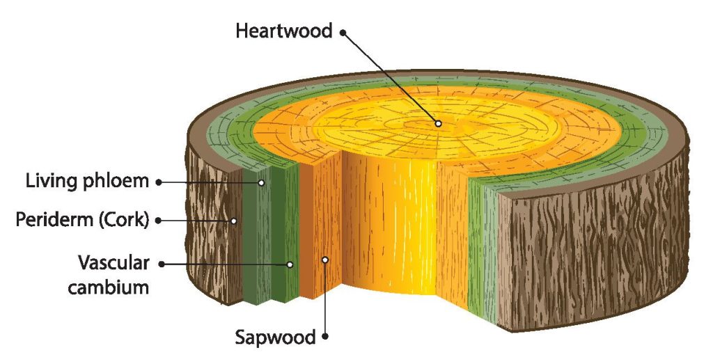
Vascular cambium first appears as a cylinder of actively dividing cells between primary xylem and primary phloem. Vascular cambium gives rise to two new tissues, one is the secondary xylem next to the inner surface of the vascular cambium, the other is the secondary phloem appearing outer to the vascular cambium.
The secondary xylem causes most of the increase in stem thickness. Over the years a woody stem gets thicker and thicker as its vascular cambium produces layer upon layer of secondary xylem. These layers are visible as rings. Since one growth ring is formed in one year, a count of the rings at the base of trunk indicates the age of a tree at the time it was cut.
In most trees, the conduction of water and dissolved substances by secondary xylem become limited to the outer or younger portion of that tissue. As trees grow older only few annual growth rings are active in conduction at one time. The active portion is called sap wood. The inactive non[1]conducting wood is called heartwood.
In most species, the heartwood accumulates a variety of chemicals such as resins, oils, gums and tannins. These provide a resistance to decay and insect attack, for example, in red cedar and conifers.
Another important function of the cambium is to form callus or wood tissue on or over the wound, soft parenchymatous tissues are rapidly formed on or below the damaged surface of stems and roots. Callus also unites the branches during budding and grafting.

(ix) What are the main differences between exoskeleton and endoskeleton.
ANSWER:
The main differences between exoskeletons and endoskeletons are:
EXOSKELETON:
(1) External to the body, covering and protecting the soft tissues and organs underneath.
(2) Typically made of non-living materials like chitin (insects and crustaceans) or calcium carbonate (shells).
(3) Fixed size, requiring molting to grow larger.
(4) Can be rigid or flexible depending on the material and structure.
(5) Examples: Insects, spiders, crustaceans, mollusks (shells).
ENDOSKELETON:
(1) Internal to the body, providing support and structure from within.
(2) Composed of living tissues, primarily bone and cartilage.
(3) Grows continuously with the organism throughout its life.
(4) More flexible due to joints and muscles attached to the bones.
(5) Examples: Vertebrates (humans, mammals, birds, reptiles, fish).
(x) List the functions of skeleton.
ANSWER:
FUNCTIONS OF SKELETON:
Some major functions of the skeletal system are as follows:
(i) Support and shape: Bones support soft tissues and serve as attachment sites for most muscles and provide shape to the body.
(ii) Protection: Bones protect critical internal organs, such as brain, spinal cord, heart and lungs.
(iii) Movement: Skeletal muscles attached to the bones help in moving the body.
(iv) Mineral homeostasis: Bones serve as store for calcium, phosphorus, sodium and potassium. Through negative feedback mechanisms, bones can release or take up minerals to maintain homeostasis.
(v) Blood cell production: Red and white blood cells are produced in bone marrow, a connective tissue found within certain bones.
(xi) Explain the role of osteoclasts in remodeling of bone and describe the structure of compact bone.
ANSWER:
Consult textbook at page 31.
(xii) List the main parts of axial skeleton.
ANSWER:
Consult textbook at page 31 — 32.
(xiii) Distinguish between fibrous, cartilaginous and synovial joints.
ANSWER:
Fibrous Joints:
These joints are held together by short collagen fibers embedded in connective tissue. Such joints are present in the skull, and they fix teeth into the jaw.
Cartilaginous Joints:
These joints allow little or no movement. Hyaline cartilage forms joint between growing bone. The bones held together by fibrous cartilage are found between vertebrae at the point where coaxal bones meet in front of the pelvis.
Synovial Joints:
These joints contain a cavity filled with fluid and are adapted to reduce friction between the moving joints. The joint is surrounded by a layer of connective tissue called “fibrous capsule” and their inner layer the synovial membrane. Some parts of capsule may be modified to form distinct ligament, holding the bones together.
(xiv) Discuss methods of locomotion in fish, land vertebrates and birds.
ANSWER:
Consult textbook at page 46 —48.
(xiv) Distinguish between the following:
(a) Axial skeleton and appendicular skeleton.
ANSWER:
Axial Skeleton:
The axial skeleton includes the skull, the vertebrae, ribs and the sternum.
Appendicular Skeleton:
The appendicular skeleton consists of pectoral girdle and appendages (fore limbs), and pelvic girdle and appendages (hind limbs).
(b) Phototactic and chemotactic stimulus.
ANSWER:
Phototactic Stimulus
“This refers to the movement in response to stimulus of light. The movement may be towards the source of light (positive) or away from the source of light (negative).”
Example: The best example of positive tactic movement is the passive movement of chloroplast due to cyclosis. This movement helps the chloroplast to absorb maximum light for CO2 fixation. The light intensity and direction both affect the intra cellular distribution of chloroplasts.
Chemotactic Stimulus
“This involves the movement in response to stimulus of chemicals or chemical gradients in the environment.”
Example: The movements shown by sperms of liver-worts, mosses, ferns towards archegonia in response to stimulus of nucleic acid released by the ovum is one such example.
(c) Osteocytes and osteoblast.
ANSWER:
Osteocytes:
Mature bone cells embedded in the bone matrix, involved in maintenance and regulation of bone tissue.
Osteoblast:
Bone-forming cells responsible for the synthesis and deposition of bone matrix during bone formation.
(d) Brachialis and brachioradialis.
ANSWER:
Brachialis:
(1) Muscle responsible for flexing the forearm at the elbow joint, located deep beneath the biceps brachii.
(2) Its origin is in humerus and insertion in ulna.
Brachioradialis:
(1) Muscle that aids in flexing the forearm at the elbow joint, with a major role in pronation and supination; runs along the forearm, connecting to the radius bone.
(2) Its origin is in humerus and insertion in radius.
(e) Origin and of muscles.
ANSWER:
Origin:
Origin is the end of muscle which remains fixed when muscle contracts.
Insertion:
Insertion is the end of the muscle that moves the bone.
(f) Bone and cartilage.
ANSWER:
Bone and cartilage are different types of connective tissues in the body.
Bone:
Bones are hard, rigid structures composed of cells, collagen, and minerals, providing support, protection, and a site for blood cell formation. They are highly vascularized and have a higher regenerative capacity.
Cartilage:
In contrast, cartilage is a flexible tissue with chondrocytes and a gel-like matrix, offering cushioning in joints and support to certain structures. Cartilage is avascular, has limited regenerative abilities, and is softer in texture compared to bones.
(g) Troponin and tropomyosin.
ANSWER:
Troponin and tropomyosin are muscle proteins that work together to regulate muscle contraction.
Troponin:
Troponin is a complex that includes subunits with specific functions related to calcium binding and actin-myosin interaction.
Tropomyosin;
Tropomyosin physically covers the myosin-binding sites on actin. It cooperates with troponin in response to calcium signals to initiate or inhibit muscle contraction.
NOTE: Please go through these solutions and give your opinion about the quality of the work. You may also give suggestions to improve the contents. Thanks!